
.gif)
European Journal of Case Reports and Clinical Images
European Journal of Case Reports and Clinical Images
Clinical Image | Open Access
Volume 2026 - 2 | Article ID 305 | http://dx.doi.org/10.51521/EJCRCI.2026.e22.119
Academic Editor: Dr. Jerry P
1Resident in
Internal Medicine Training, Department of Internal Medicine, Unidade Local de
Saúde do Oeste – Hospital Caldas da Rainha, Caldas da Rainha, Portugal; ORCID:
https://orcid.org/0000-0001-9503-7049.
2Resident in Internal Medicine Training, Department of Internal
Medicine, Unidade local de Saúde do Alto Alentejo – Hospital de Portalegre,
Portalegre, Portugal, ORCID: https://orcid.org/0000-0002-1994-7849.
3Senior Consultant in Internal Medicine, Casa Santa Maria
(RNCCI/ERPI), Lisboa, Portugal; ORCID: https://orcid.org/0000-0003-4448-4169
Corresponding Author: Sara Lourenço Tereso, ORCID: https://0000- 0001-9503-7049,
Rua Diário de Notícias, 2500-176, Caldas da Rainha, Portugal.
Citation: Sara Tereso, Elisabete Mendes, Carlos Machado e Costa
(2026). Expansive Maxillary Brown Tumor in a Patient with Secondary
Hyperparathyroidism. Euro J Case Rep Clin Imag. 2026; August, e22,1-3.
Copyrights: © Sara Lourenço Tereso, 2026, et al., This article is
licensed under the Creative Commons Attribution-Non
Commercial-4.0-International-License-(CCBY-NC)
(https://europeanjournalofcasereports.com/blogpage/copyright-policy). Usage and
distribution for commercial purposes require written permission.
Image Case Presentation:
A
31-year-old woman from Angola, with chronic kidney disease of unknown aetiology
and on a haemodialysis programme since 2015, presented to the emergency
department with a progressively enlarging and deforming nasopalatine mass that
caused dysphagia and impaired vision in the left eye. Laboratory tests showed
negative infectious serologies, Parathyroid Hormone (PTH) of 3462 pg/ml,
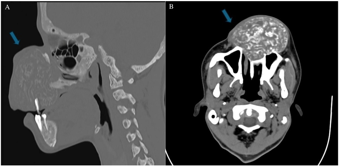
phosphate 4.1 mg/dl and calcium 9.2 mg/dl. Maxillofacial computed tomography
revealed a 78 × 60 × 63 mm mass in the anterosuperior maxillary region, with
diffuse alteration of bone density in the context of a disordered phosphocalcic
metabolism. Biopsy demonstrated fibrovascular tissue, multinucleated giant cells
and haemosiderin deposits, consistent with a brown tumour (Figures 1A, 1B). A
diagnosis of brown tumour secondary to secondary hyperparathyroidism was
established. The patient underwent parathyroidectomy, with a subsequent
decrease in PTH to 348 pg/ml.
Brown tumour
is an uncommon osseous manifestation of secondary hyperparathyroidism,
typically seen in chronic kidney disease, particularly in patients undergoing
haemodialysis [1,2]. Although more frequent in women, craniofacial involvement
is rare [1,2]. It is characterised by medullary osteofibrosis and increased
focal osteoclastic bone resorption leading to fibrous osteitis [1]. Its
expansive behaviour and radiological appearance may mimic malignant lesions
[1]. Diagnosis requires clinical, biochemical, radiological and histological
correlation, and parathyroidectomy is the treatment of choice to correct the
underlying metabolic disorder and promote regression of the lesions [1,3].
Declarations:
Funding: This
research received no external funding.
Institutional Review Board Statement: Not applicable.
Informed Consent Statement: Informed consent for publication was obtained from the patient’s
legal guardians.
Data Availability Statement: No new data were created or analyzed in this study. Data sharing
is not applicable to this article.
Acknowledgments: Not applicable.
Conflicts of Interest: We confirm that there are no conflicts of interest to declare.
References:
1. Shavlokhova V, Goeppert B, Gaida MM, Saravi B, Weichel F, Vollmer A,
et al. Mandibular brown tumor secondary to hyperparathyroidism: a case report
with five-year follow-up and literature review. Int J Environ Res Public
Health. 2021 July,18(14):7370. DOI: 10.3390/ijerph18147370.
2. Lacativa PG, Franco FM, Pimentel JR,
Patrício PJ, Filho GMD, Farias ML. Prevalence of radiological findings among
cases of severe secondary hyperparathyroidism. Sao Paulo Med J. 2009 May,
127(2):71–77. DOI: 10.1590/s1516-31802009000200020004.
3. Panagopoulos A, Tatani I, Kourea HP, Kokkalis ZT, Panagopoulos K, Megas P.
Osteolytic lesions (brown tumors) of primary hyperparathyroidism misdiagnosed
as multifocal giant cell tumor of the distal ulna and radius: a case report. J
Med Case Rep. 2018 June, 12(1):176. DOI: 10.1186/s13256-018-1723-y.


.png)








.jpg)


.png)