
.gif)
European Journal of Case Reports and Clinical Images
European Journal of Case Reports and Clinical Images
Clinical Image | Open Access
Volume 2026 - 2 | Article ID 308 | http://dx.doi.org/10.51521/EJCRCI.2026.e22.122
Academic Editor: John Bose
Corresponding Author: Elisabete Dulce da Cunha Mendes, Internal Medicine Resident,
Medical Degree, Department of Internal Medicine, Unidade Local de Saúde do
Norte Alentejano, Hospital de Portalegre, Portalegre, Portugal, ORCID:
http://orcid.org/0000-0002-1994-7849.
Citation: Elisabete Dulce da Cunha Mendes, Sara Lourenço Tereso,
Rita Afonso Diz (2026). Gastric Gastrointestinal Stromal Tumor with Pancreatic
Invasion: A Rare Presentation. Euro J Case Rep Clin Imag. 2026; August,
e22,1-3.
Copyrights: © Elisabete Dulce da Cunha Mendes, 2026, et al., This
article is licensed under the Creative Commons Attribution-Non
Commercial-4.0-International-License-(CCBY-NC)(https://europeanjournalofcasereports.com/blogpage/copyright-policy).
Usage and distribution for commercial purposes require written permission.
Keywords: Gastrointestinal Stromal Tumour, GIST, Gastric Tumour, Pancreatic
Invasion, Computed Tomography.
Image Case Presentation:
Gastrointestinal
stromal tumours (GISTs) are mesenchymal neoplasms that most commonly arise in
the stomach and may occasionally reach an exceptionally large size and invade
adjacent organs. Their management depends on tumour extent, metastatic disease
and molecular characteristics, with tyrosine kinase inhibitors playing a
central role in advanced disease [1-3].
We present
the case of a 65-year-old woman who presented with several weeks of nausea,
vomiting and significant weight loss. Physical examination revealed a large,
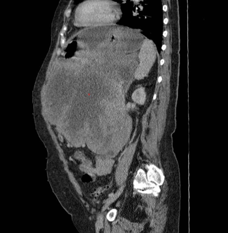
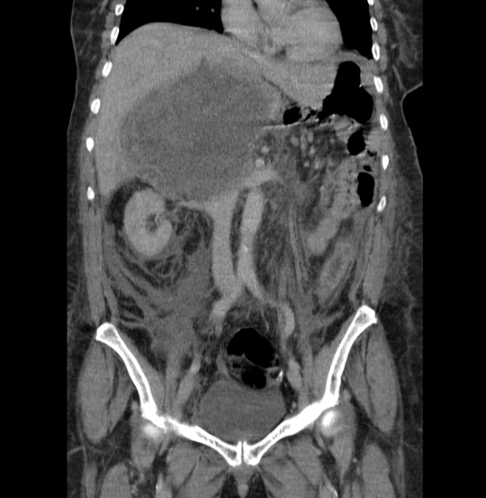
painless abdominal mass. Computed tomography (CT) demonstrated a heterogeneous
intra-abdominal mass measuring approximately 28 cm, with extensive central
necrosis and peripheral enhancing soft tissue, displacing the stomach, spleen,
bowel and other abdominal structures; the pâncreas could not be clearly
delineated (Figure 1).
The patient
underwent extensive en bloc resection, including near-total gastrectomy, distal
pancreatectomy, splenectomy, left colectomy and cholecystectomy.
Histopathological examination demonstrated a 30-cm high-grade gastric GIST with
pancreatic invasion (pT4N0M1R1). Immunohistochemistry was positive for CD34,
CD117 and DOG1, and three peritoneal metastatic nodules were identified.
Imatinib therapy was initiated. Two years later, CT demonstrated extensive
recurrent disease, with a 20 × 10 cm partially necrotic subhepatic mass
invading the liver parenchyma and hepatic hilum (Figure 2).The recurrence was considered unresectable and the patient was referred
for palliative care. This case illustrates the potential for gastric GISTs to
reach an enormous size, invade adjacent organs and recur despite treatment.
Cross-sectional imaging is essential for defining tumour extent, assessing
resectability and monitoring disease progression.
Declarations:
Funding: This
research received no external funding.
Institutional Review Board Statement: Not applicable.
Informed Consent Statement: Not applicable.
Data Availability Statement: No new data were created or analyzed in this study. Data sharing is
not applicable to this article.
Acknowledgments: Not applicable.
Conflicts of Interest: The authors declare no conflict of interest.
References:
1. Casali
PG, Blay JY, Abecassis N, et al. Gastrointestinal stromal tumours:
ESMO-EURACAN-GENTURIS Clinical Practice Guidelines for diagnosis, treatment and
follow-up. Ann Oncol. 2022;33(1):20–33.
2. von
Mehren M, Kane JM, Riedel RF, et al. NCCN Guidelines® Insights:
Gastrointestinal Stromal Tumors, Version 2.2022. J Natl Compr Canc Netw.
2022;20(11):1204–1214. DOI:10.6004/jnccn.2022.0058.
3. Serrano C, Álvarez R, Carrasco JA, et al. SEOM-GEIS clinical
guideline for gastrointestinal stromal tumors (2022). Clin Transl Oncol.
2023;25(9):2707–2717. DOI:10.1007/s12094-023-03177-7.


.png)








.jpg)


.png)