
.gif)
European Journal of Case Reports and Clinical Images
European Journal of Case Reports and Clinical Images
Clinical Image | Open Access
Volume 2026 - 2 | Article ID 309 | http://dx.doi.org/10.51521/EJCRCI.2026.e22.123
Academic Editor: John Bose
1Resident in
Internal Medicine Training, Department of Internal Medicine, Unidade Local de
Saúde do Oeste – Hospital Caldas da Rainha, Caldas da Rainha, Portugal; ORCID:
https://orcid.org/0000-0001-9503-7049.
2Resident in
Internal Medicine Training, Department of Internal Medicine, Unidade local de
Saúde do Alto Alentejo – Hospital de Portalegre, Portalegre, Portugal, ORCID:
https://orcid.org/0000-0002-1994-7849.
3Hospital Assistant
in Internal Medicine, Department of Internal Medicine, Unidade local de Saúde
de Santa Maria – Hospital de Santa Maria, Lisboa, Portugal.
4Senior Consultant
in Internal Medicine, Casa Santa Maria (RNCCI/ERPI), Lisboa, Portugal; ORCID:
https://orcid.org/0000-0003-4448-4169.
Corresponding Author: Sara Lourenço Tereso, ORCID: https://orcid.org/0000-0001-9503-7049, Rua Diário de Notícias, 2500-176, Caldas da Rainha, Portugal.
Citation: Sara
Tereso, Elisabete Mendes, Tânia Vassalo, Carlos Machado e Costa (2026).
Incomplete Left Third Cranial Nerve Palsy of Microvascular Etiology. Euro J
Case Rep Clin Imag. 2026; August, e22,1-3.
Copyrights: © Sara Lourenço Tereso, 2026, et al., This article is licensed under the Creative Commons Attribution-Non Commercial-4.0-International-License-(CCBY-NC) (https://europeanjournalofcasereports.com/blogpage/copyright-policy). Usage and distribution for commercial purposes require written permission.
Image Case
Presentation:
A 71‑year‑old man with uncontrolled
hypertension and dyslipidemia presented to the emergency department with
binocular diplopia, more pronounced at near, and left eyelid ptosis. He denied
headache, ocular pain, jaw claudication, visual acuity changes, or other
neurological symptoms. On examination, he had isocoric and reactive pupils,
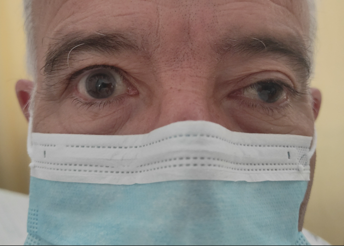
left eyelid ptosis, left eye exo‑hypotropia (Figure 1), horizontal diplopia on
dextroversion and vertical diplopia on infraversion, with limitation of
adduction and infraduction. The remaining neurological examination was
unremarkable. Laboratory tests showed no relevant abnormalities. Cranial CT and
CT angiography revealed no parenchymal lesions, extra‑axial collections, or
aneurysms. During hospitalization, he remained hemodynamically stable, with
normalization of blood pressure after starting ramipril. Etiological workup
showed negative autoimmune and infectious screening, HbA1c 5.7% and LDL‑c 108
mg/dL. Brain MRI demonstrated hyperintensities in the corona radiata and
bilateral centrum semiovale, consistent with chronic microvascular disease,
without compressive lesions along the oculomotor pathway. Neurology diagnosed
incomplete third cranial nerve palsy of probable microvascular etiology.
Diplopia resolved during hospitalization, with residual ptosis. At neuro‑ophthalmology
follow‑up, he had normal ocular motility, without diplopia or ptosis.
Third cranial nerve palsy may result from compressive, infectious,
inflammatory, or microvascular ischemic causes [1], with incidence increasing
with age [2] and peaking in the 70s and 90s [2]. In adults with vascular risk
factors, microvascular etiology is the most frequent, particularly when the
pupil is spared [1]. Exclusion of aneurysm, neoplasm, and head trauma is
mandatory [1–4]. Prognosis is generally favorable, with complete recovery
within weeks after strict control of hypertension and dyslipidemia [1].
Keywords: Oculomotor Nerve Palsy, Microvascular Ischemia, Diabetes Mellitus, Arterial Hypertension.


.png)








.jpg)


.png)