
.gif)
European Journal of Case Reports and Clinical Images
European Journal of Case Reports and Clinical Images
A Case Report | Open Access
Volume 2025 - 1 | Article ID 230 | http://dx.doi.org/10.51521/EJCRCI.2025.e11.106
Academic Editor: John Bose
Julia Murlewska�, Krzysztof Serafin2, Iwona
Strzelecka1,3, Maria Respondek-Liberska1,3
�
1Department of Prenatal Cardiology Polish Mother's Memorial
Hospital, Research Institute, ?�d?, Poland.
2Department of Gynaecology and Obstetrics, Faculty of
Medicine, Jagiellonian University Medical College, Krakow, Poland.
3Department for Fetal Malformations Diagnoses &
Prevention Medical University of Lodz, Faculty of Public Health.
�
Corresponding author: Julia Murlewska, MD, PhD, Department of Prenatal Cardiology, Polish Mother's Memorial Hospital Research Institute, Rzgowska 281/289, 93-338 ?�d? Poland. e-mail: juliamurlewska.jm@gmail.com
Citation: Julia Murlewska, Krzysztof Serafin,
Iwona Strzelecka, Maria Respondek-Liberska (2025). Recovery of Segmental
Ventricular Contractility in a Fetus with Mixed Arrhythmia: Monitoring with
Fetal Speckle-Tracking Echocardiography. Euro J Case Rep Clin Imag. 2025; Nov, e11,
1-5.
Copyrights � 2025, Julia Murlewska. et al., This
article is licensed under the Creative Commons
Attribution-Non-Commercial-4.0-International-License-[CCBY-NC]
[https://europeanjournalofcasereports.com/blogpage/copyright-policy]. Usage and
distribution for commercial purposes require written permission. Nursing,
Nagpur, India
Abstract:
Background: Fetal arrhythmias can disrupt
coordinated myocardial contraction and, in severe cases, impair cardiac
function. While conventional echocardiography assesses global systolic and
diastolic performance, subtle segmental disturbances may go undetected. Fetal
speckle-tracking echocardiography (STE) provides quantitative evaluation of
ventricular mechanics and may offer added value in monitoring therapy response.
Case
Presentation: A
30-year-old pregnant woman at 27+3 weeks� gestation was referred for suspected
fetal arrhythmia. She had occupational exposure to various chemicals but no
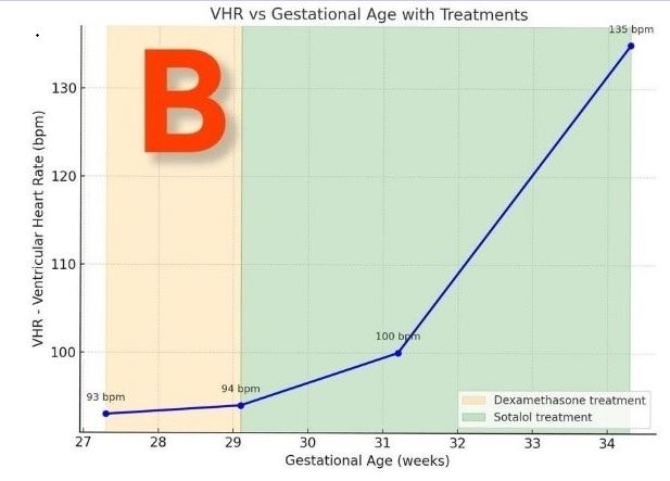
autoimmune antibodies. Fetal echocardiography revealed ventricular bradycardia
(93 bpm) with atrial rate of 160 bpm, prolonged atrioventricular conduction
with intermittent Wenckebach-type block, frequent atrial and ventricular
ectopy, and cardiomegaly (CTAR 0.40).
STE (FetalHQ) demonstrated global
segmental left ventricular (LV) dysfunction and right ventricular (RV)
impairment except in basal segments, despite no signs of hydrops. Maternal
therapy with intravenous dexamethasone and oral salbutamol was initiated. Serial
follow-up showed gradual normalization of ventricular rate, regression of
cardiomegaly, and progressive recovery of segmental function, culminating in
complete restoration of RV contractility and near-normal LV performance by 34+3
weeks.
Conclusions: This case highlights the utility of fetal STE in detecting regional ventricular dysfunction and objectively documenting myocardial recovery in fetuses with mixed arrhythmias. Integration of advanced imaging into routine surveillance may improve assessment of treatment efficacy, guide management, and provide reassurance to both clinicians and parents when rhythm stabilization leads to functional recovery.


.png)


.jpg)


.png)