
.gif)
European Journal of Case Reports and Clinical Images
European Journal of Case Reports and Clinical Images
Clinical Image | Open Access
Volume 2026 - 2 | Article ID 306 | http://dx.doi.org/10.51521/EJCRCI.2026.e22.120
Academic Editor: Dr. Jerry P
Corresponding Author: Elisabete
Dulce da Cunha Mendes, Internal Medicine Resident, Medical Degree, Department
of Internal Medicine, Unidade Local de Saúde do Norte Alentejano, Hospital de
Portalegre, Portalegre, Portugal, Email: vitoria.cantim@gmail.com ; ORCID:
http://orcid.org/0000-0002-1994-7849.
Citation: Elisabete Dulce da Cunha Mendes, Sara Lourenço Tereso,
Rita Afonso Diz (2026). The Real Culprit Behind the Cannonball Pattern. Euro J
Case Rep Clin Imag. 2026; August, e22,1-3.
Copyrights: © Elisabete Dulce da Cunha Mendes, 2026, et al., This
article is licensed under the Creative Commons Attribution-Non
Commercial-4.0-International-License-(CCBY-NC)
(https://europeanjournalofcasereports.com/blogpage/copyright-policy). Usage and
distribution for commercial purposes require written permission.
Image Case
Presentation:
The “cannonball” pattern
describes the radiological appearance of multiple, well-defined, rounded
pulmonary nodules of variable size. It is a characteristic manifestation of
haematogenous pulmonary metastases, although the differential diagnosis
includes infectious, inflammatory and vascular conditions [1]. Renal cell
carcinoma is classically associated with this appearance, while colorectal and
other solid malignancies may also present with multiple “cannonball” metastases
[1–3].
We present an 87-year-old
woman with colon adenocarcinoma who presented to the emergency department with
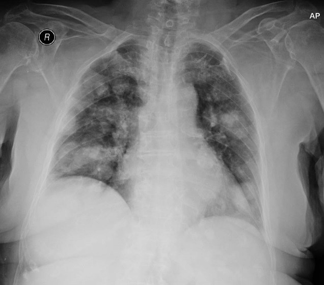
worsening dyspnoea and several days of disorientation. Chest radiography
revealed multiple bilateral pulmonary nodules in a diffuse “cannonball” pattern
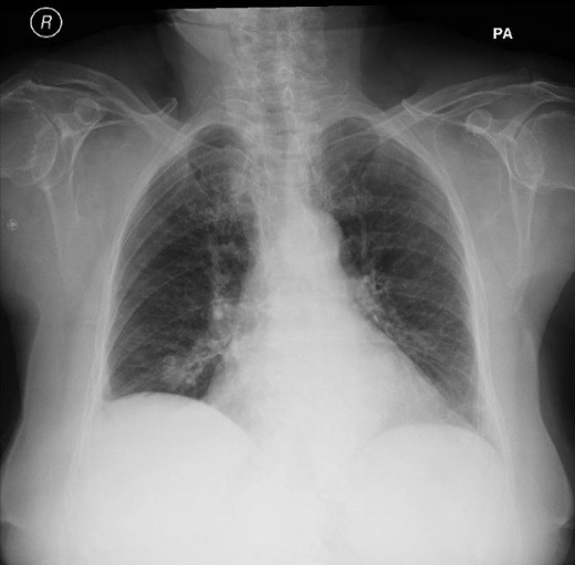
(Figure 1). Remarkably, these findings were absent on a chest radiograph
obtained less than one year earlier (Figure 2), highlighting the rapid
progression of the disease. Brain computed tomography demonstrated a 2.8-cm
rounded, heterogeneous lesion in the deep left temporoparietal region, adjacent
to the ventricular system, suspicious for a neoplastic lesion. Further
investigation confirmed pulmonary and cerebral metastases from colon
adenocarcinoma.
This case highlights the
importance of recognising the “cannonball” pattern as a radiological clue to
metastatic disease, particularly in patients with a known malignancy.
Comparison with previous imaging may provide an important clue to disease
progression and help guide further diagnostic evaluation and management.
Keywords: Pulmonary Metastases, Pulmonary Nodules, Chest Radiography, Cannonball Pattern.


.png)








.jpg)


.png)