
.gif)
European Journal of Case Reports and Clinical Images
European Journal of Case Reports and Clinical Images
Clinical Image | Open Access
Volume 2025 - 1 | Article ID 292 | http://dx.doi.org/10.51521/EJCRCI.2025.e11.108
Academic Editor: John Bose
Corresponding author: Miguel Bernardino Antunes Vicente, E-mail: miguelantunesv@gmail.com; Orcid: https://orcid.org/0000-0002-1329-5229, Praceta Professor Mota Pinto, 3004-561, Coimbra, Portugal.
Citation: Miguel Vicente, Gonçalo Batista
(2025). Vieussens and Kugel Coronary Rings in a Patient with a Lesion of the
Distal Left Main Coronary Artery. Euro J Case Rep Clin Imag. 2025; Dec, e11,
1-2.
Copyrights: © Miguel Vicente, 2025 This
article is licensed under the Creative Commons
Attribution-Non-Commercial-4.0-International-License-(CCBY-NC)
(https://europeanjournalofcasereports.com/blogpage/copyright-policy). Usage and
distribution for commercial purposes require written permission.
Imaging Case
An 87-year-old man with hypertension, dyslipidemia, diabetes, and a
history of smoking presented to the emergency department with mild chest pain
radiating to the left upper limb for two days. The pain occurred after
unusually intense physical exertion, did not improve with rest, and had
worsened in the preceding hours.
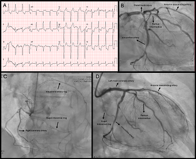
On admission, the electrocardiogram (Figure 1A) showed ischemic changes,
and high-sensitivity troponin was elevated (22,000 ng/L). Transthoracic
echocardiography revealed diffuse hypokinesia, apical akinesia, and a reduced
left ventricular ejection fraction (35%).
Coronary angiography (Figure 1B) demonstrated a 90% distal left main
coronary artery stenosis involving the left anterior descending (LAD), ramus
intermedius, and circumflex (Cx) arteries. The LAD showed diffuse disease with
a 90% stenosis in the mid-segment; the dominant Cx had a critical 90% proximal
stenosis. The right coronary artery (RCA) was small-caliber, with a 90%
proximal stenosis. Collateral circulation through the Vieussens
arterial ring and Kugel’s interatrial ring (Rentrop
3) was visualized (Figure 1C).
The Vieussens arterial ring, first described by Raymond
Vieussens in the 17th century, and Kugel’s interatrial ring,
identified by Maurice Kugel in 1927, are rare anatomic variants with estimated
prevalences of 3% and 6%, respectively. These structures connect the conal
branch of the RCA to branches of the left main coronary artery and may be
present even in the absence of coronary artery disease. However, they acquire
clinical significance when providing effective collateral flow between the left
and right coronary circulations in the setting of severe coronary stenosis.
In this case, the collateral circulation supplied additional perfusion to the
LAD and Cx, allowing the patient to remain asymptomatic for a prolonged period
despite severe lesions. Percutaneous coronary intervention was performed on the
stenotic lesions (Figure 1D), requiring rotational atherectomy. Due
to intra-procedural hypotension, aminergic support was
necessary.
''See article image in pdf''


.png)


.jpg)


.png)